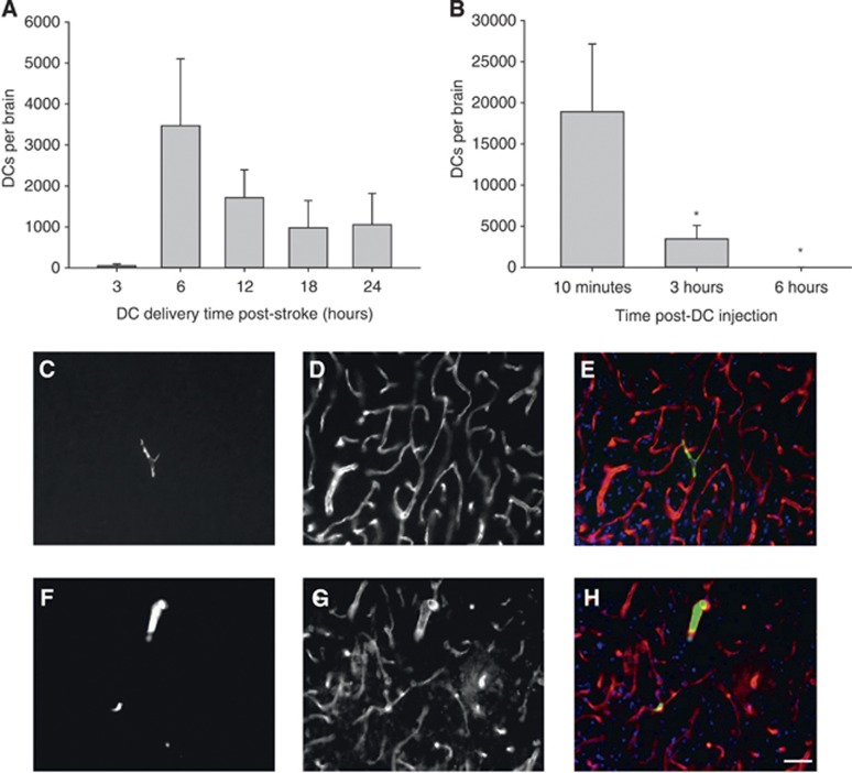
Figure 1.
(A) Animals subjected to transient middle cerebral artery occlusion (tMCAO) were injected with green fluorescent protein (GFP+) ex vivo-derived dendritic cells(exDCs) at 3 (N=4), 6 (N=7), 12 (N=5), 18 (N=6) and 24 hours (N=5) hours poststroke, then killed 3 hours postinjection. The total number of exDCs in serial sections spanning the stroke region is indicated and error bars indicate s.e.m. (B) Animals subjected to tMCAO were injected with exDCs at 6 hours poststroke and killed at 10 minutes (N=7), 3 hours (N=7), or 6 hours (N=6) postinjection. One-way analysis of variance and S–N–K post hoc revealed that the number of exDCs at 10 minutes is significantly higher than 3 hours (P=0.043) and 6 hours postinjection (P=0.05). Error bars indicate s.e.m. (C–H) Animals injected with GFP+ exDCs (green) at 6 hours poststroke were killed at 10 minutes (N=2) or 3 hours (N=2) poststroke. Forty-micrometer-thick serial slices were stained for the endothelial marker RECA-1 (red) and counterstained with the nuclear marker 4',6-diamidino-2-phenylindole I (DAPI) (blue). (C) Representative section of 10 minutes poststroke GFP+ exDC (D) RECA-1 stain (E) overlay with DAPI (N=2 animals, 69 total cells counted). (F) Representative section of 3 hours poststroke GFP+ exDC (G) RECA-1 stain (H) overlay with DAPI (N=2 animals, 50 cells counted). Scale bar 20 μm.

