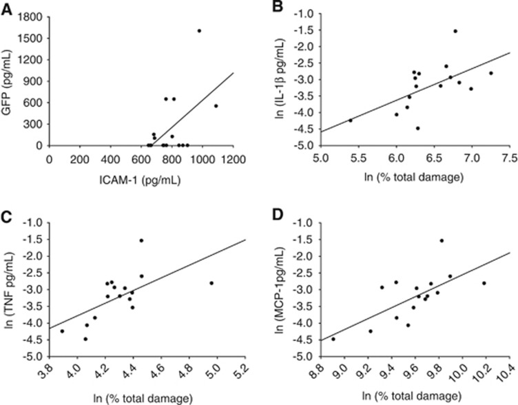
Figure 3.
Animals subjected to transient middle cerebral artery occlusion were injected with green fluoroscent protein (GFP+) ex vivo-derived dendritic cells at 6 hours post-stroke and killed 10 minutes postinjection. Two millimeter slices were taken and slices one, three, and five were analyzed for protein, both sides of slices two and four were analyzed for stroke damage using 2,3,5-triphenyltetrazolium chloride. Brain homogenate was run on a GFP ELISA and 11-plex polystyrene bead inflammatory protein assay. (A) GFP ELISA and ICAM-1 multiplex values were significantly correlated (R2=0.291, P=0.0314). (B) Total damage was significantly correlated with IL-1β (R2=0. 353, P=0.0152), (C) TNF (R2=0.386, P=0.0102), and (D) MCP-1 (R2=0.454, P=0.00421) after log-linear analysis.

