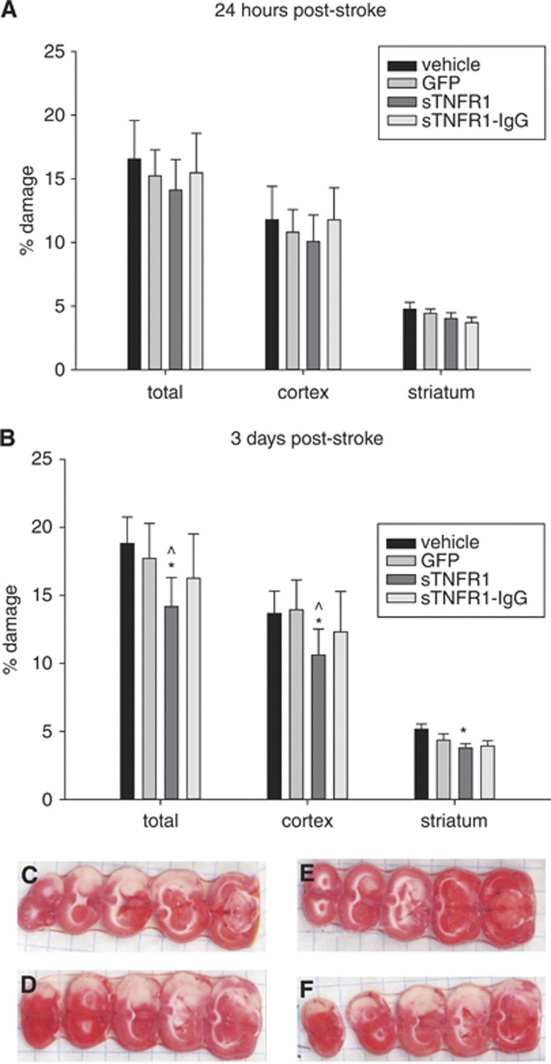
Figure 5.
(A) No significant differences in damage at 24 hours poststroke. (B) At 3 days poststroke, there was a significant reduction in percent total (P=0.008), percent cortex (P=0.039), and percent striatum (P=0.006) versus vehicle damage, and versus green fluorescent protein (GFP)-ex vivo-derived dendritic cell treated animals in percent total (P=0.038) and percent cortex (P=0.044) damage *P<0.05 in 2-way analysis of variance (ANOVA) (group, slice) for each brain area compared with vehicle; ^P<0.05 in 2-way ANOVA (group, slice) for each brain area compared with GFP control. 24 hours vehicle (N=14), GFP (N=16), sTNFR1 (N=14), sTNFR1-IgG (N=11) and 3 days vehicle (N=16), GFP (N=13), sTNFR1 (N=13), sTNFR1-IgG (N=9). Error bars indicate s.e.m.. Representative photos of 2,3,5-triphenyltetrazolium chloride staining for day 3 infarct size assessment for (C) vehicle, (D) GFP-exDCs, (E) sTNFR1-exDCs, (F) sTNFR1-IgG-exDCs.

