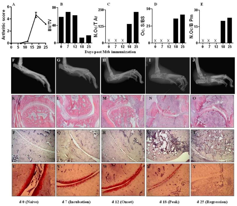
Figure 1. Progression of arthritic inflammation and tissue damage in the hind paws of Lewis rats during the course of AA.

First (Top) panel: (A), Arthritic scores of rats beginning from d 0 (Mtb injection) through d 25; (B-E) Histomorphometric analysis of bone tissue: (B) Bone volume versus total tissue volume (BV/TV), (C) number of osteoclasts per tissue area (N.Oc/T Ar), (D) active resorption per bone surface area based on the ratio of osteoclast surface/bone surface area (Oc.S/BS), and (E) number of osteoclasts per bone perimeter (N.Oc/B Pm). Second panel (F-J), Representative radiographs of hind limbs at different phases of the disease course. Third panel (K-O), Representative micrographs of H&E-stained histological sections (10×) of the hind paw joints showing joint space, pannus containing the mononuclear cell infiltrate, and bone mass at different phases of the disease course. Fourth panel (P-T), representative micrographs of the stained histological sections showing TRAP-positive cells (20×); Fifth (Bottom) panel (U-Y), representative micrographs of the safranin O’-stained histological sections showing proteoglycan content in the cartilage (20×). Mtb, heat-killed M. tuberculosis H37Ra; X- parameter tested but not detectable.
