
Figure 2. Use of cultured human fibroblasts as a target to assay for donor-specific antibodies.


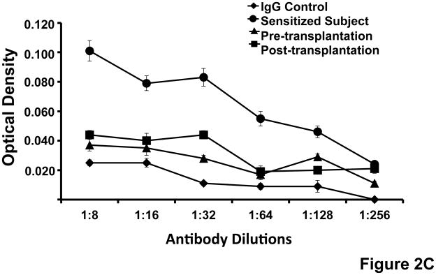
(A)Expression of HLA-class I on cultured human fibroblasts. The figure shows that cultured fibroblasts express HLA-class I detected by binding of a murine monoclonal antibody specific for a non-polymorphic domain of HLA–A, B and C (W6/32) and measured measured by ELISA. Standard errors of triplicate measurements are indicated. (B) Flow cytometry analysis of HLA-class I and class II expression by cultured fibroblasts. Shown is the light scatter plot with the lymphocyte gate depicted in red. Dot plots show staining with antibody isotype control, anti-HLA class I and anti-HLA class II antibodies. The fractions of positively stained cells are noted. (C)Use of cultured fibroblasts for assay of donor-specific antibodies in the serum of renal transplant recipients. Cultured donor fibroblasts were incubated sequentially with serial dilutions of serum from a highly sensitized subject and with serum obtained before and two months after renal transplantation and with AP labeled anti-human IgG. Donor-specific IgG was measured by detecting the optical density at 405 nm (Y-axis). The assay detects binding of IgG from a highly sensitized subject but minimal or no binding of IgG from the renal transplant recipient or pooled IgG used a control. Standard errors of triplicate measurements are indicated.