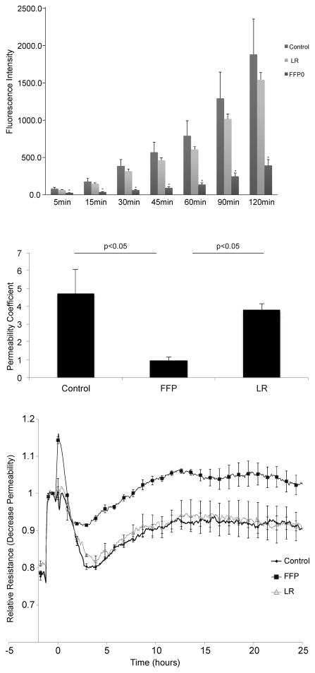
Figure 1. FFP decreases pulmonary endothelial cell permeability in vitro.
A. Fluorescence intensity over time in a transwell endothelial cell permeability assay for cells treated with 10% LR or FFP diluted in basal medium. At all time points, fluorescence in FFP treated cells was significantly lower than in LR and control treated cells (*=p<0.05), while there was no significant difference at any time point between LR and controls. B. Permeability coefficients for each group at 10% concentration. All permeability coefficients are expressed as cm/min. Error bars represent the standard error of the mean. C. TEER measured at 4000 hertz in cells treated with 10% FFP, 10% LR or control media. FFP significantly increased transendothelial resistance compared to controls and LR (p < 0.05 by ANOVA with Tukey post hoc tests) indicating decreased endothelial permeability. Each trace represents the mean trace from three replicates. Dots appear with error bars indicating standard error of the mean at 1 hour intervals.

