Figure 4.
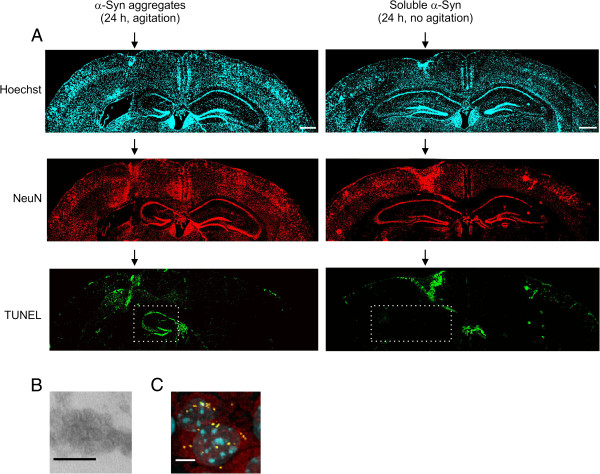
In vivo neurotoxicity of synthetic α-Syn oligomers. (A) α-Syn protein was treated as indicated and injected into the brain of wild-type C57BL/6 mice. Apoptotic neurons of the hippocampal region adjacent to the injection site were detected by TUNEL assay (green channel) followed by confocal microscopy. Hippocampus in the injected hemisphere is shown in a dotted box. The neuronal marker NeuN is detected by immunofluorescence (red channel). Nuclei are stained with Hoechst (blue channel). Scale bar, 500 μm. (B) Electron micrographs of toxic α-Syn oligomers obtained after incubation at 37°C for 24 h. Scale bar, 25 nm. (C) Synthetic α-Syn aggregates were labeled with Alexa Fluor 633 prior to injection and detected by confocal microscopy (aggregates, yellow channel; NeuN, red channel; nucleus, blue channel). Scale bar, 10 μm.

