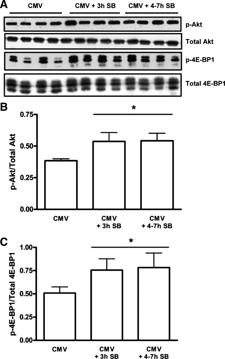
Fig. 7.
A: representative immunoblots for phosphorylated Akt (p-Akt), total Akt, phosphorylated eukaryotic initiation factor 4E binding protein (p-4E-BP1), and total 4E-BP1. B: mean densitometric values for the amount of activated Akt. Values are shown as the ratio of p-Akt to total Akt, from CMV (n = 7), CMV + 3 h SB (n = 6), and CMV + 4–7 h SB (n = 7) diaphragms, measured on the same blotting membrane, and are expressed as a percentage of CMV. B: mean densitometric values for the amount of p-4E-BP1. Values are shown as the ratio of p-4E-BP1 to total 4E-BP1, from CMV (n = 7), CMV + 3 h SB (n = 6), and CMV + 4–7 h SB (n = 7) diaphragms, measured on the same blotting membrane. Values are means ± SD. *P < 0.01, CMV + 3 h SB and CMV + 4–7 h SB vs. CMV.

