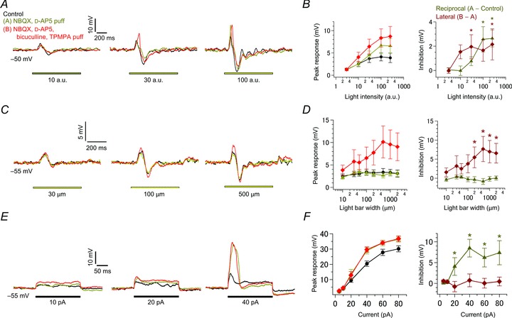
Figure 6. Pharmacological separation of reciprocal and lateral inhibition.

A, effects of light intensity. Voltage responses in an intact terminal (Vm: ∼−50 mV; current-clamped condition) to various intensities of light (yellow bar; 500 μm in width) were recorded in control (black), in the presence of puff-applied 10 μm NBQX and 20 μm d-AP5 (yellow; selective suppression of reciprocal inhibition), and in the presence of puff-applied 100 μm BIC and 800 μm TPMPA in addition to NBQX and d-AP5 (red; suppression of reciprocal and lateral inhibition). ECl was −70 mV. B, the relationship between the peak response amplitude and light intensity in each condition as shown in A (left; n= 9). Activation of reciprocal (dark yellow) and lateral inhibition (dark red) was estimated as the difference between responses in each condition (right). C and D, effects of light bar width as in A and B (n= 10–12 in D). Voltage responses in an intact terminal (Vm: ∼−55 mV; current-clamped condition) to various widths of light (yellow bar; intensity of 100 a.u.) were recorded in each condition as shown in A (C). ECl was −70 mV. E and F, effects of current injection as in A and B (n= 8–11 in F). Voltage responses to current injection (black bar) were recorded from an intact terminal (Vm: ∼−55 mV; current-clamped condition)in each condition as shown in A (E). ECl was −70 mV. BIC, bicuculline; d-AP5, d-(–)-2-amino-5-phosphonopentanoic acid; NBQX, 2,3-dioxo-6-nitro-1,2,3,4-tetrahydrobenzo[f]quinoxaline-7-sulphonamide; TPMPA, (1,2,5,6-tetrahydropyridin-4-yl) methylphosphinic acid.
