
Figure 3. Adenosine release is evoked by glutamate receptor agonists.
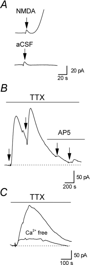
A, focal application of NMDA (0.5 s puff) onto the surface of CA1 induced a small pressure artefact (at arrow) followed by a much larger, delayed slower component which is absent if only aCSF is applied (using the same perfusion pipette at the same pressure). B, focal application of NMDA (0.5 s puff) induced an increase in the extracellular concentration of adenosine in the presence of 1 μm TTX to block action potentials. NMDA was applied twice in control (1 μm TTX, application at arrows) and then was applied in the presence of the NMDA receptor antagonist d-AP5 (50 μm), which prevented an increase in adenosine concentration. This confirms that increases in adenosine concentration evoked by puffing on NMDA result from NMDA receptor activation rather than from a pressure change. C, superimposed biosensor adenosine traces following application of glutamate (1 s puff) in control aCSF (with 1 μm TTX) and in Ca2+-free aCSF (3 mm Mg2+, 1 mm EGTA). Glutamate was applied in Ca2+-free aCSF first, to prevent any run down of release due to depletion of adenosine stores, and then an increase in adenosine concentration was evoked after normal aCSF was washed in.