
Figure 10. Scheme of adenosine release in the hippocampus.
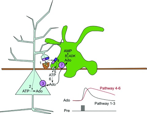
Glutamate release from the Schaffer collaterals releases glutamate, which can activate NMDA and AMPA receptors on the postsynaptic spine (1) of a pyramidal neuron and also NMDA and AMPA receptors on nearby astrocytic processes (4). By some means, yet to be elucidated, this activation of glutamate receptors increases the metabolic load on the neuron and leads to an increase in intracellular adenosine (2), which is then rapidly transported out of the cell by the ENTs (3). In parallel with this, the activation of glutamate receptors on the astrocyte (4) leads to an increase in intracellular Ca2+ and increased exocytosis of ATP-containing vesicles (5). Once in the extracellular space, ATP is broken down to adenosine (6). Extracellular adenosine can be taken up into astrocytes via ENTs (7) as the astrocytes maintain very low intracellular adenosine levels as a consequence of the activity of adenosine kinase (ADK), which converts adenosine to AMP (8). The two parallel pathways have differing kinetics: the neuronal pathway of direct release (1–3) has rapid kinetics, while those of the indirect astrocytic pathway (4–6) are slower.