
Figure 2. Response of a representative NI neuron to icv CRF in vivo that was RLN3-immunopositive.
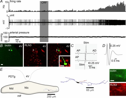
A, application of icv 3 μg CRF (grey shaded column) significantly increased the discharge rate of the neuron by ∼350%, with onset of effect at 130 s post-infusion. There was little change in arterial blood pressure following icv CRF infusion. B, immunofluorescent visualization of the recorded and juxtacellularly labelled neuron revealed it was RLN3-positive. C, four superimposed traces in which a spontaneous spike (AP) triggered an electrical stimulus in the medial septum (Stim; mean 10.91 ± 0.06 ms) that elicited a constant latency antidromic spike (AD; upper trace; mean 9.30 ± 0.11 ms). Spike collision (lower trace) when the stimulation was applied within the critical interval (mean 9.02 ± 0.04 ms), indicates that this neuron projects to the medial septum. D, the recorded neuron had a short spike duration of 0.63 ms. E, light microscopic reconstruction (63× magnification) of a biotinamide-labelled RLN3 neuron that was activated by CRF. The location of the neuron is within the boundaries of the NI. Soma (black), dendrites (blue) and an axon (red) are drawn from five sections of 50 μm thickness. Juxtacellular labelling revealed the neuron expressed RLN3 (inset). Scale bar in photomicrographs of B= 20 μm and E= 15 μm. CRF, corticotrophin-releasing factor; 4V, fourth ventricle; icv, intracerebroventricular; mlf, medial longitudinal fasciculus; NI, nucleus incertus; NIc, NI pars compacta; NId, NI pars dissipata; PDTg, posterodorsal tegmental nucleus.