
Figure 3. Response of a representative NI neuron to icv CRF in vivo that was RLN3-immunonegative.
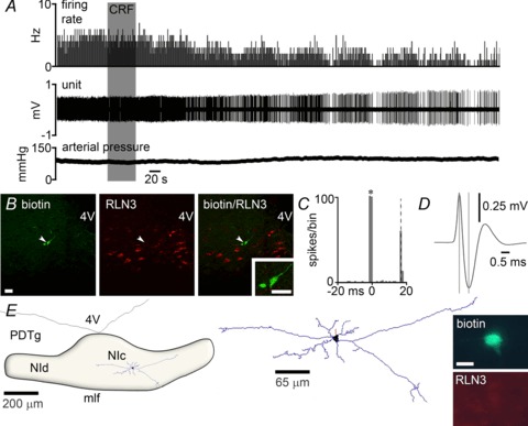
A, application of icv 3 μg CRF (grey shaded column) significantly decreased the discharge rate of the neuron by ∼50%, with onset of effect at 140 s post-infusion. Arterial blood pressure increased by ∼15 mmHg following icv CRF infusion. B, immunofluorescent visualization of the recorded and juxtacellularly labelled neuron revealed it was RLN3-negative. C, peri-stimulus time histogram (100 sweeps) of orthodromic stimulation of firing elicited by medial septum stimulation (*), indicates that the recorded neuron receives excitatory projections from the medial septum, with a delay of ∼17 ms. D, the recorded neuron had a short spike duration of 0.60 ms. E, light microscopic reconstruction (63× magnification) of a biotinamide-labelled non-RLN3 neuron that was inhibited by CRF. The location of the neuron is within the boundaries of the NI. Soma (black), dendrites (blue) and an axon (red) are drawn from six sections of 50 μm thickness. Juxtacellular labelling revealed the neuron lacked RLN3 (inset). Scale bar in photomicrographs of B= 20 μm and E= 15 μm. CRF, corticotrophin-releasing factor; 4V, fourth ventricle; icv, intracerebroventricular; mlf, medial longitudinal fasciculus; NI, nucleus incertus; NIc, NI pars compacta; NId, NI pars dissipata; PDTg, posterodorsal tegmental nucleus.