
Figure 5. CRF excitation of NI neurons in vitro.
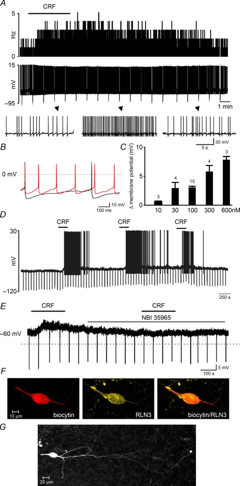
A, a representative trace of current clamp recording demonstrating the effects of bath-applied CRF (100 nm, horizontal bar) on the activity of an NI neuron, shown as a change in firing frequency (1 s bins, upper trace) and corresponding raw signal (lower trace) with amplified segments below. B, overlay of the raw signal recorded from the CRF-activated neuron before (black) and after (red) CRF treatment, revealing an increase in action potential firing frequency and decrease in afterhyperpolarization peak and duration after CRF treatment. C, dose-dependent change in membrane potential following CRF-induced depolarization. Values are presented as means ±s.e.m. and the number of neurons tested for each concentration is indicated on each bar. D, current clamp recording demonstrating the effect of three consecutive CRF applications (100 nm, horizontal bars) illustrating the sustained sensitivity of NI neurons to multiple CRF stimulations. E, membrane potential depolarization of an NI neuron following 100 nm CRF application in the presence of tetrodotoxin citrate (1 μm), and a failure to depolarize to a second application of CRF applied in the presence of the CRF receptor 1 antagonist NBI 35965 (1 μm, lower horizontal bar). F, confocal projection image (1 μm optical slice) of a recorded, CRF-activated neuron confirmed that it was RLN3-positive. G, photomicrograph of a biocytin-filled RLN3 neuron in the NI pars compacta of a 250 μm brain slice. CRF, corticotrophin-releasing factor; NI, nucleus incertus.