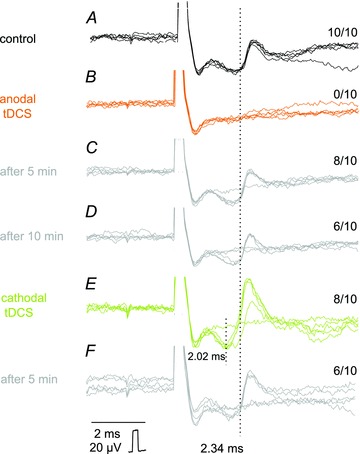
Figure 4. Opposite effects of anodal and cathodal transcranial polarization on MLF-evoked unitary EMG responses.

A–F, individual records from a neck muscle evoked by single 30 μA stimuli (five superimposed records). These were gathered before tDCS (control), during the positive or the negative tDCS and at the end of the following 5 min periods, as indicated to the left. All-or-none appearance of these potentials indicates that they most likely represented EMG responses of single motor units (in A–D) or two motor units (in E and F) and the rate of their appearance could therefore be counted (figures to the right are for 10 single records of the unit in A). It can be seen that positive tDCS prevented activation of EMG responses evoked at latencies of about 2.34 ms and the negative tDCS ensured a regular appearance of these responses and in addition activation of two other motor units, one at the same and another at a shorter latency. Dotted lines indicate the onset of the shorter and longer EMG potentials. Note that after the end of tDCS the responses tended to return to the approximately pre-tDCS level. In this and the following figures the negativity is upward and the largest shock artefacts are truncated.
