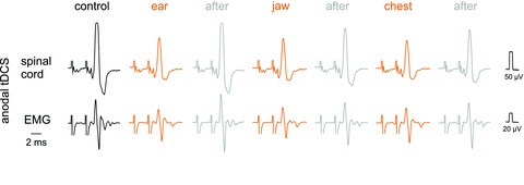
Figure 9. Qualitatively similar effects of tDCS at different locations of the reference electrode.

Upper records in each panel are from the surface of the spinal cord in the C1 segment (as in Fig. 5) while lower records are from a neck muscle (as in Fig. 4). The responses were evoked by 27 μA stimuli applied in the MLF. Anodal tDCS was applied through an electrode on the right side of the skull just rostral to the bregma with the reference electrode in contact with the left ear lobe, the lower jaw, or the chest, as indicated. Note that all responses evoked during tDCS were smaller than those evoked either before or after the tDCS periods. The illustrated responses were evoked by the 3rd stimulus, the earlier parts of the records and the shock artefacts having been cropped off.
