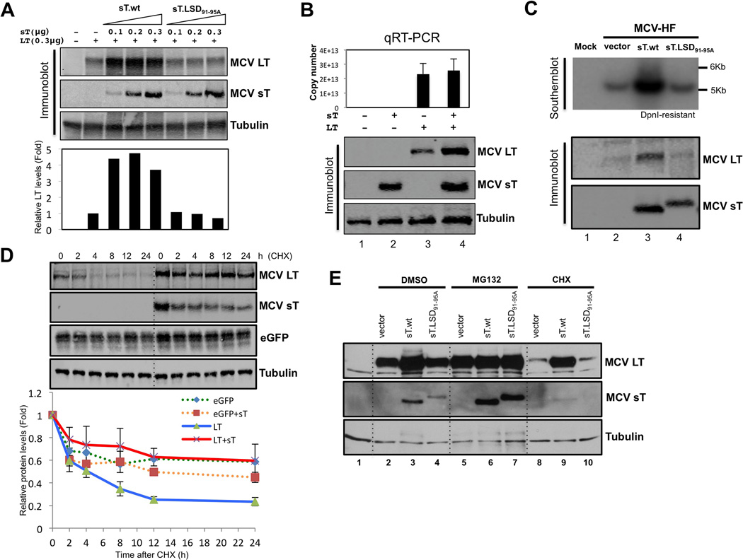
Figure 3. MCV sT inhibits proteasomal degradation of MCV LT.
(A) MCV sT expression increases LT expression through a domain encompassed by amino acids 91–95. Extremly low levels of wild type small T (sT.wt) expression (0.1 µg transfected plasmid) results in fully saturated (5–7 folds) LT stability. In contrast, the titration of mutant (sT.LSD91-95A) expression, even over a broad range of levels, has no effect on LT stability. Quantitative LICOR immunoblotting for LT expression was determined using CM2B4 antibody in triplicate, with a representative shown. (B) MCV sT expression does not transcriptionally regulate MCV LT expression. 293 cells were transfected with empty vector (lane 1, negative control), sT alone (lane 2), LT alone (lane 3), LT and sT together (lane 4), and quantitative RT-PCR was performed to detect LT mRNA transcription. Corresponding levels of sT and LT protein expression by immunoblotting are shown. Error bars represent SEM; n = 3. (C) sT expression in trans increases MCV replication. MCV-HF (0.3 µg) was cotransfected with empty vector (lane 2), sT wild type (lane 3), sT.LSD91-95A mutant (lane 4) (0.3 µg each) into 293 cells and replication was assayed by Southern blotting. Wild type MCV sT cotransfection markedly activates viral replication (upper panel) and LT expression (lower panel) from the MCV-HF clone, whereas the MCV sT LSD mutant does not. (D) MCV sT specifically inhibits LT protein turnover. LT protein turnover was measured by a cycloheximide (CHX) chase assay using quantitative immunoblot analysis. LT (0.3 µg) and eGFP (0.3 µg) constructs were cotransfected together with either empty vector or sT plasmid (0.3 µg). Cells were treated with CHX (0.1 mg/ml) 24 hours after transfection and harvested at each time point indicated. Protein expression was quantified in triplicate using an LI-COR IR imaging system (bottom). Coexpression of sT extended the half-life of LT from ~3–4 h up to >24 h, but did not significantly affect eGFP protein turnover. Error bars represent SEM; n = 3. .(E) MCV sT inhibits proteasomal degradation of LT protein. 293 cells were transfected with LT with either wild-type sT or sT.LSD91-95A. Cells were treated with MG132 (10 µM) or CHX (0.1 mg/ml) 24 h after transfection. Comparable levels of LT accumulation are seen with empty vector or sT.LSD91-95A expression to those seen with wild-type sT expression during MG132 treatment. See also Figure S2

