Fig. 2.

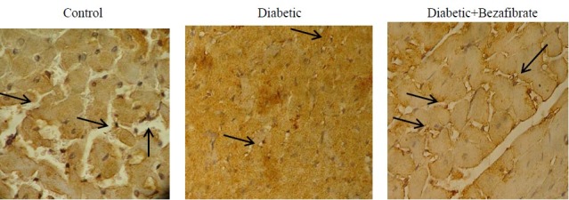
Representative photographs of immunohistochemical staining (×400) of myocardial tissue with anti-CD31 monoclonal antibody in experimental groups. Arrows indicate CD31 positive cells.

Representative photographs of immunohistochemical staining (×400) of myocardial tissue with anti-CD31 monoclonal antibody in experimental groups. Arrows indicate CD31 positive cells.