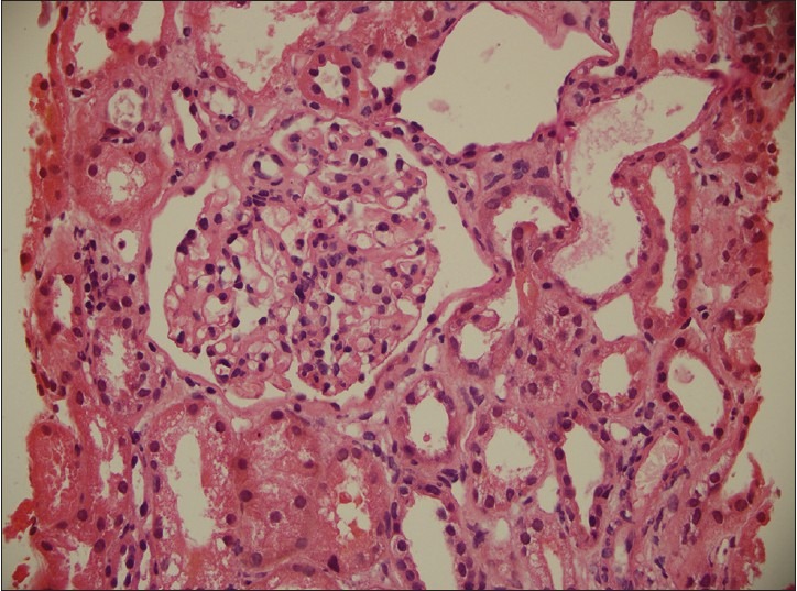
Figure 1.

There is mild increase in mesangial matrix with mesangial hypercellularity, mild thickening of the paramesangial capillary walls with narrowing of capillary lumina. The extraglomerular compartment showed focal mild interstitial fibrosis, focal tubular dilatation with flattening of lining epithelium, hyaline casts, focal infiltrates of lymphocytes and occasional eosinophils
