FIGURE 2.
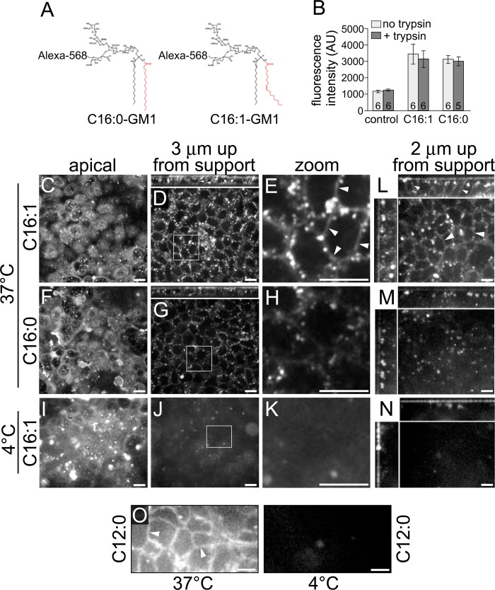
Polarized epithelial cells sort GM1 gangliosides into the basolaterally directed transcytotic pathway on the basis of the structure of the ceramide domain. A, schematic of GM1 structures. B, cell lysates of MDCK monolayers preincubated with the indicated Alexa Fluor-labeled GM1 species were measured by fluorimetry and reported as arbitrary units (AU) of fluorescence (± S.E.). The number of monolayers/condition is indicated at the column base. C–O, polarized epithelial monolayers were incubated apically with the indicated fluorophore-labeled GM1 species at 10 °C for 1 h, shifted to either 37 °C or 4 °C for the times indicated below, and imaged live by confocal microscopy. C–K, MDCK monolayers were incubated for 2 h at the indicated temperatures and imaged at the indicated Z plane (small panels above D and G show X-Z reconstructions). Zoomed images (E, H, and K) are from insets in D, G, and J. L–O, T84 monolayers were incubated apically with the indicated GM1 species for 3 h at the temperatures shown. Main panels in L-N are X-Y optical sections imaged 2 μm up from the basolateral support, and small panels at the top and left are X-Z and Y-Z reconstructions, respectively. O, T84 monolayers were incubated with Alexa Fluor-conjugated C12:0-GM1 at the indicated temperature and imaged 2 μm up from the basolateral support. Arrowheads indicate basolateral membrane labeling in E, L, and O. Scale bars = 10 μm. Data are representative of three independent experiments for B and O and at least six independent experiments for C–N.

