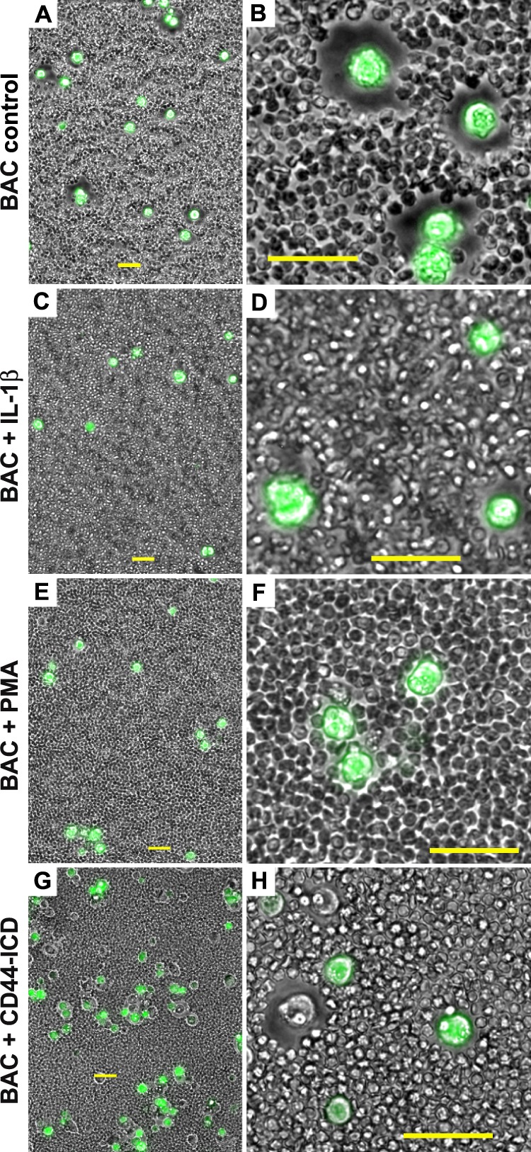
FIGURE 1.

Conditions that promote CD44 cleavage alter the chondrocyte pericellular matrix. Pericellular matrices were visualized by the exclusion of formalin-fixed erythrocyte particles from the cells surface. Bovine articular chondrocytes (BAC) were incubated in the absence (A and B) or presence of 10 ng/ml IL-1β (panels C and D) or 10 mm PMA (E and F). Images shown in panels A--F represent two-color overlays of phase contrast and green fluorescence images, green fluorescence due to calcein-AM that stains live cells and was used to provide contrast. In panels G and H, bovine articular chondrocytes were co-transfected with CD44-ICD-myc and empty pEGFP-N2 plasmid. Images shown in G and H represent two-color overlays of phase contrast and green fluorescence due to EGFP. As such, successfully transfected bovine articular chondrocytes are GFP-positive and express the CD44-ICD; non-transfected cells within the same field are GFP-negative and serve as controls. Panels A, C, E, and G are lower magnification views to allow visualization of multiple cells; panels B, D, F, and H are higher magnification views to highlight details. All bars are 50 μm.
