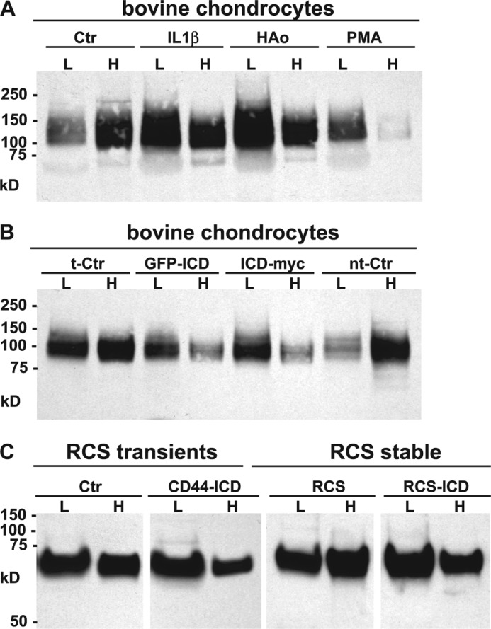
FIGURE 2.

Differential extraction of full-length CD44 from chondrocytes. High density monolayers of bovine articular or RCS chondrocytes were treated under a variety of conditions and then detergent extracted using the sequential differential extraction approach (0.1% Igepal, lanes labeled as L; 0.5% Igepal and 1% Empigen, lanes labeled H). Equal-volume aliquots were analyzed by Western blotting using the CD44 anti-cytotail antibody for detection. Panel A is a representative experiment of bovine chondrocytes treated without or with IL-1β, HA oligosaccharides (HAo) or PMA. Panel B is a representative experiment of bovine chondrocytes that were transfected with a control plasmid (t-Ctr), N-terminal GFP-tagged CD44-ICD (GFP-ICD), C-terminal myc-tagged CD44-ICD (ICD-myc), or non-transfected control chondrocytes (nt-Ctr). On these Western blots, full-length CD44 of bovine chondrocytes is represented by a diffuse band between 95 and 150 kDa. Panel C is a representative experiment depicting control RCS cells or RCS cells transiently or stably transfected with CD44-ICD. Full-length CD44 of RCS chondrocytes is represented by a diffuse band between 65–85 kDa.
