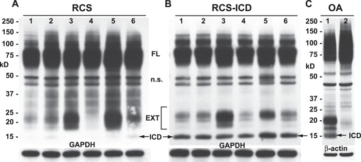
FIGURE 4.

Changes in CD44 protein after stable transfection of RCS cells with CD44-ICD. High density monolayers of parental rat chondrosarcoma chondrocytes (panel A) or RCS cells stably transfected with CD44-ICD (panel B) were treated under a variety of conditions. After treatment, the cells were lysed, and aliquots of equivalent protein were analyzed by Western blotting using the anti-CD44 cytotail antibody for detection. Depicted are untreated control cells (lanes 1) or cells treated with IL-1β (lanes 2), IL-1β + DAPT (lanes 3), IL-1β + GM6001 (lanes 4), PMA (lanes 5), or IL-1β + MG132 (lanes 6). Bands represent full-length CD44 (FL), nonspecific bands (n.s.), CD44-EXT fragments (EXT), and CD44-ICD fragments (ICD). After detection of CD44, the Western blots were reprobed for GAPDH. The CD44 profile of a human osteoarthritic (OA) chondrocyte lysate is shown for comparison (panel C). Lysates derived from cells grown in monolayer (lane 1) or alginate beads (lane 2) were immunoblotted using anti-CD44 cytotail antibody. Arrows in all panels are used to further highlight CD44-ICD bands.
