FIGURE 7.

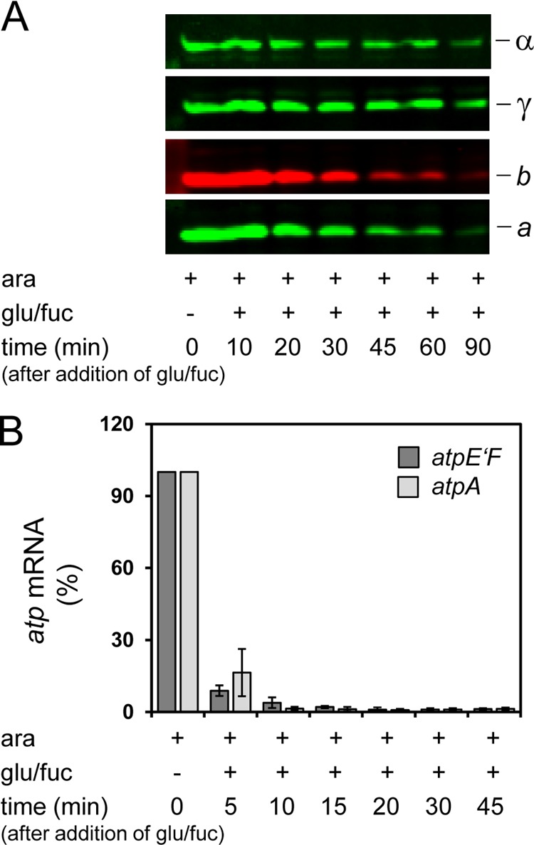
Stability of FOF1−δ (A) and degradation of atp mRNA (B) after repression of ParaBAD controlling expression of atpBEFH*AGDC. DK8 carrying plasmids pBAD33.Δδ, pET22.atpH-TTG and pT7POL26 (FOF1−δ) was grown as described under “Experimental Procedures.” At each time point indicated, cells were harvested for immunoblot analysis (A) and isolation of RNA (B). A, stability of FOF1−δ. After resuspension of cell lysates in sample loading buffer, cells were incubated for 5 min at 99 °C. The amount of cell extract (20 μg/lane) was calculated according to the determination of Neidhardt et al. (34) that 160 μg of protein is present per ml of growth medium at OD = 1. Proteins were separated by SDS-PAGE and detected by immunolabeling as indicated. B, degradation of atp mRNA. rt-RT-PCR was performed using primer pairs atpE′F (dark gray) and atpA (light gray). The amount of atp mRNA present in the FOF1 sample grown with Ara was set to 100%. Error bars, S.E.
