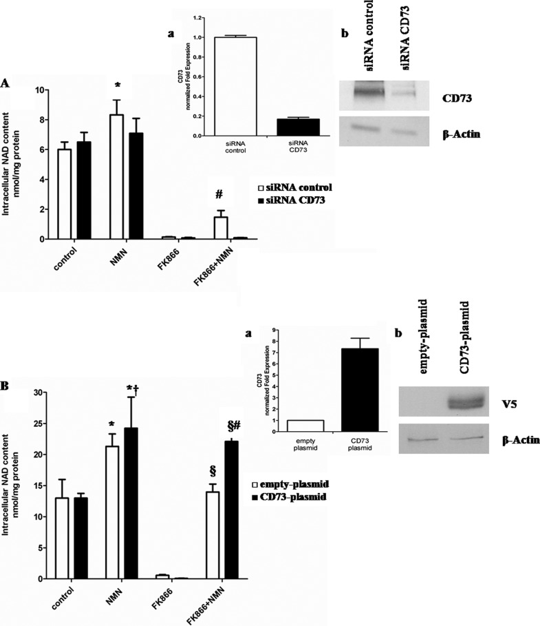
FIGURE 4.
CD73 expression affects intracellular NAD+ synthesis triggered by extracellular NMN. A, A549 cells were transfected with specific siRNA for CD73 or with negative control (siRNA control) and seeded in 12-well plates (2 × 10⁵ cells/well). Cells were treated for 72 h with or without 30 nm FK866 in the presence or absence of 10 μm of NMN (added twice a day). Cells were harvested and lysed in 0.6 m PCA, and NAD+ content was measured in neutralized extracts. NAD+ values were normalized to protein content. Data are expressed as mean ± S.D. (error bars) (n = 3). *, p < 0.05 compared with untreated siRNA control cells; #, p < 0.01 compared with FK866-treated siRNA control cells and with FK866 + NMN-treated siRNA CD73 cells. Inset, 24 h after transfection; a, qPCR analysis was performed, and expression of CD73 was normalized to that of the housekeeping genes GAPDH and HPRT1 and compared with negative control; b, Western blot analysis of CD73 protein level was performed (results from one representative experiment are shown). B, U87 cells were transfected in parallel with pcDNA3.1/V5-HisTOPO® (empty plasmid) or with CD73-pcDNA3.1/V5-His TOPO® (CD73 plasmid) using the Nucleofector system and seeded in 12-well plates (2 × 10⁵ cells/well). Cells were then treated for 72 h with or without 30 nm FK866 in the presence or absence of 10 μm of NMN (added twice a day). Cells were harvested and lysed in 0.6 m PCA, and NAD content was measured in neutralized extracts. NAD+ values were normalized to protein content. Data are expressed as mean ± S.D. (n = 3). *, p < 0.05 compared with untreated empty plasmid cells; †, p < 0.05 compared with untreated CD73 plasmid cells; §, p < 0.0001 compared with FK866-treated empty plasmid cells; #, p < 0.01 compared with FK866 + NMN-treated empty plasmid cells. Inset, 24 h after transfection; a, transfected cells were subjected to total RNA extraction, and qPCR analysis for CD73 was performed. Results are normalized on the reference genes GAPDH and HPRT1 and compared with empty plasmid; b, Western blot analysis of cell lysates was performed, using a monoclonal antibody against the V5 epitope (one representative experiment of three is shown).

