
FIGURE 5.
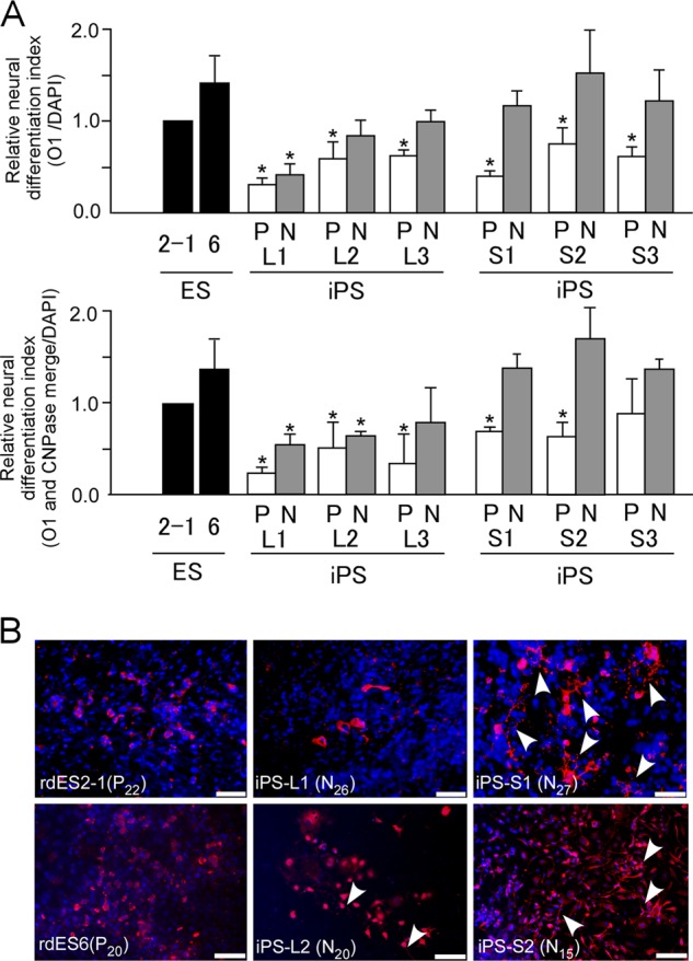
In vitro differentiation to oligodendrocytes of rabbit pluripotent stem cells after naive-like conversion. A, relative neural differentiation indices are shown for oligodendrocytes marked by O1 (left graph) and the merged signals for O1 and CNPase (right graph) derived from rabbit ES cells (rdES2-1 and rdES6) or primed state (P) and naive-like state (N) rabbit iPS cells (liver-derived iPS-L1, -L2, and -L3 cells and stomach-derived iPS-S1, -S2, and -S3 cells). Data are shown as the mean ± S.D. (rdES, n = 3; each rabbit iPS cell line, n = 3). *, p < 0.05. B, immunocytochemical detection of oligodendrocytes with ramified branches (arrowheads) using anti-O1 antibody (red) in in vitro-differentiated rabbit ES cells and naive-like-state iPS cells.