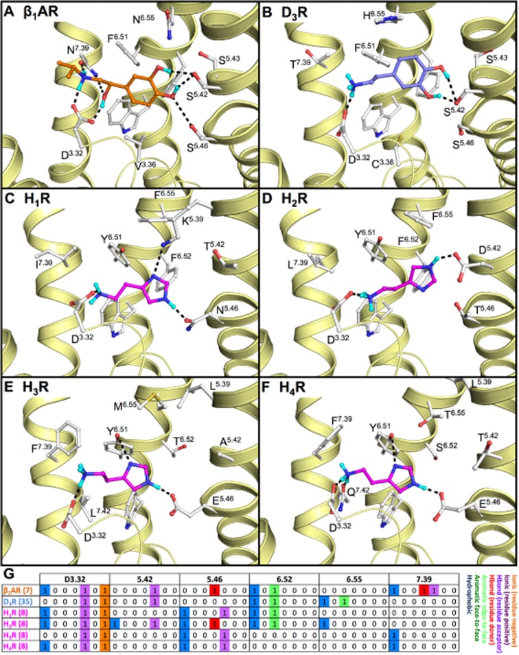
Figure 5.

Binding mode of (A) isoproterenol (7, orange carbon atoms) in the turkey β1AR (PDB code 2Y03 (Warne et al., 2012)). Proposed binding modes of endogenous agonists, based on SDM studies, of (B) dopamine (35, blue carbon atoms) in the human D3R and the proposed binding modes of histamine (8, magenta carbon atoms) in the different human histamine receptors; (C) H1R, (D) H2R, (E) H3R and (F) H4R. Minor changes in the H1R crystal structure (i.e. rotation of K1915.39 and N1985.46) allow for accommodation of histamine. The H2R, H3R and H4R models are based on the H1R crystal structure. Rendering and colour-coding are the same as in Figure 2. (G) Molecular interaction fingerprint (IFP) bit strings describing the binding poses of 7–8 and 35 (A–F), encoding different interaction types with D3.32, 5.42, 5.46, 6.52, 6.55 and 7.39 (colour-coding as described in Figure 2). Two-dimensional structures of the molecules are presented in Figure 3A and B.
