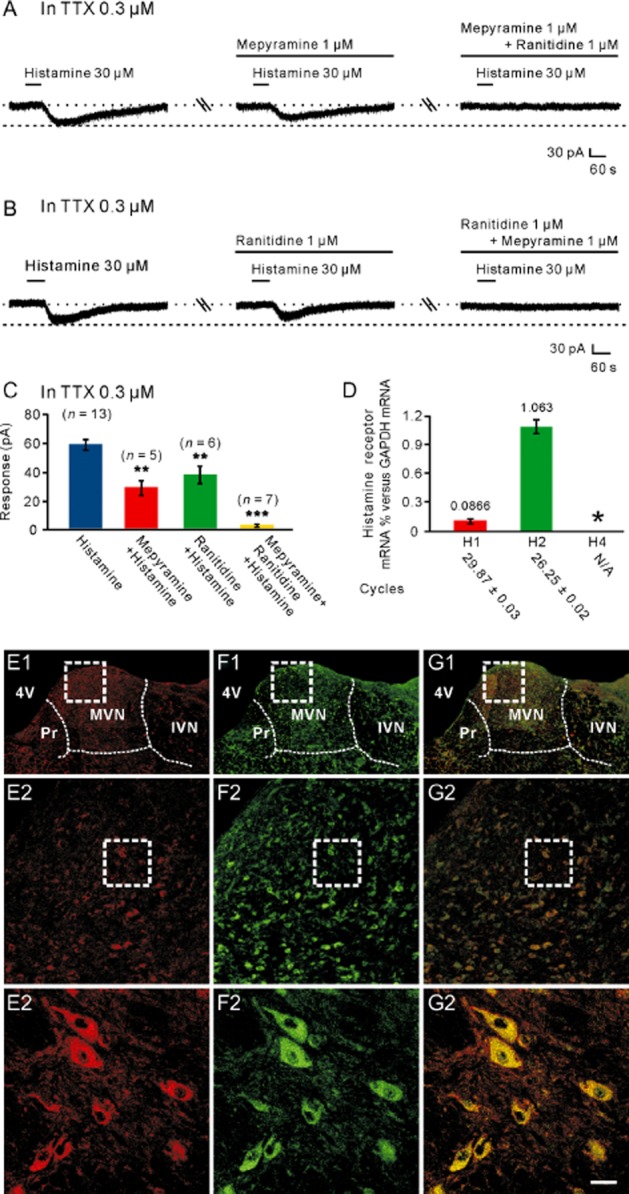
Figure 2.

H1 and H2 receptors co-mediated the histamine-induced postsynaptic excitation on MVN neurons. (A) In the presence of TTX, the histamine-induced inward currents were partly blocked by mepyramine, a highly selective H1 receptor antagonist, and abolished by combined application of mepyramine and ranitidine, a highly selective H2 receptor antagonist. (B) In the presence of TTX, the histamine-induced inward currents were partly blocked by ranitidine, and abolished by the combination of mepyramine and ranitidine. (C) Group data of the tested MVN neurons. Data shown are means ± SEM; **P < 0.01, ***P < 0.001, significantly different from control. (D) Bar graphs showing the relative expression of H1, H2 and H4 receptor mRNAs in the MVN. In this and the following figures, asterisks indicate samples showing no specific signal, and cycles necessary to get a signal are listed below each gene. (E1–G3) Double immunostaining results showed that histamine H1 (E1–E3) and H2 (F1–F3) receptors were not only present in the rat MVN but also co-localized in the same MVN neurons (G1–G3). Scale bars: (C1), (D1) and (E1), 550 μm; (C2), (D2) and (E2), 100 μm; (C3), (D3) and (E3), 20 μm. 4V, 4th ventricle; IVN, inferior vestibular nucleus; MVN, medial vestibular nucleus; Pr, prepositus nucleus.
