
Figure 3.
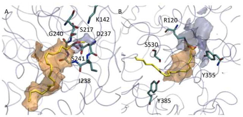
Hydrophilic (light blue) and hydrophobic (orange) isocontour surfaces of FAAH-1 (A) and COX-2 (B). For the sake of clarity, relevant residues are highlighted as stick models with C atoms colored in cyan. The protein is shown as transparent cyan tube. Substrates are methyl arachidonyl fluorophosphonate in FAAH and arachidonic acid in COX-2.