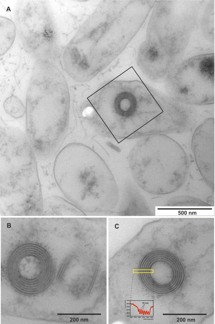
Figure 2.
Ultrastructure of L. alexandrii DFL-11T and its R-bodies. (A) Survey view of the cells from the near-surface position of a colony. Many bacterial remnants are visible, one of which contains an R-body; such bodies are shown enlarged in (B) and (C). (B) A pair of R-bodies, oriented at right angle towards each other, one as a cross-section and the other one cut oblique-longitudinally. The bipartite, black-white organization of the spiral layers is shown, and the averaged intensity profile (C, inset) of the boxed area shows a regular spacing of 10 nm.

