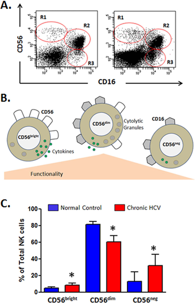
Fig. 4. Natural killer (NK) cell subset distribution is altered in HCV infection.
The expression patterns of CD56 and CD16 can identify three distinct NK cell subsets CD56brightCD16neg (R1), CD56dimCD16pos (R2), and CD56negCD16pos (R3). Representative flow cytometric dot plots of CD3neg lymphocytes (low forward and side scatter) from one normal control subject and one chronic HCV patient are shown (A). CD56bright NK cells do not display significant natural cytotoxicity and are focused on production of cytokines such as IFN-γ. This subset is considered less mature and can give rise to the CD56dim mature effector cells which are strongly cytolytic and characterized by high perforin-containing granule content. Although CD56dim NKs produce less cytokine than their CD56bright counterparts, because they represent the predominant NK subset in circulation, they are the main contributor to overall cytokine levels. The CD56negCD16pos NK subset is highly dysfunctional, has impaired cytotoxic function and poor cytokine production, and appears to be terminally differentiated (B). The bar chart shows the NK cell subset distribution which is altered in HCV infection. Median values and interquartile range are shown for normal control subjects (n = 5) compared to chronic HCV patients (n = 5). CD56dim mature effector NK cells account for the majority of circulating NK cells in both groups. Decreased CD56dim and increased CD56bright is a consistent finding in several chronic HCV cohorts (79, 87). Increased circulating levels of dysfunctional CD56negCD16pos are also evident. *p<0.05 calculated using a two sided Mann Whitney test.

