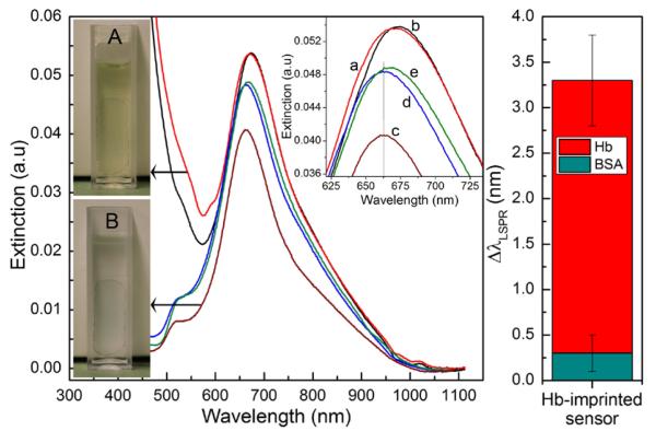
Figure 8.
Application of AuNR-imprinted sensors for the detection of hemoglobin in a real urine sample. (A) Image of the sensor substrate during incubation in urine sample. (a) and (b) represent respectively the extinction spectra of the sensor immersed in cuvette (A) before and immediately after addition of 30 μg/mL hemoglobin. After incubation in urine sample without or with the presence of target proteins, the sensor is washed and immersed in water (cuvette B) for LSPR analysis. The graphs (c-e) represent respectively the extinction spectra of the sensor substrate after incubation in normal urine (c) or after incubation in urine sample containing either BSA (d) or hemoglobin (e). The diagram on the right depicts the shift in the maximum wavelength of the Hb-imprinted nanorods after exposition to urine containing either Hemoglobin or BSA.

