Fig. 3.
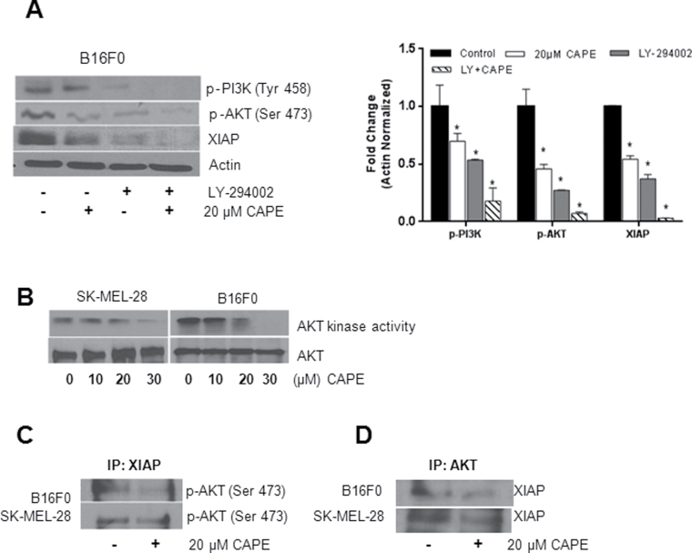
Effect of CAPE on AKT kinase activity and the interaction of AKT/XIAP in melanoma cells. (A) B16F0 cells were pretreated for 1 h with LY294002 followed by 20 µM CAPE treatment for 48 h. Cell lysates were immunoblotted with p-PI3K (Tyr 458), p-AKT (Ser 473) and XIAP. Same blots were stripped and re-probed for actin to ensure equal protein loading. *P < 0.05, statistically significant compared with control. (B) SK-MEL-28 or B16F0 cells were treated with various concentrations of CAPE for 48 h. Cell lysates were prepared and AKT kinase activity was determined using a kit according to the manufacturer’s instruction. B16F0 and SK-MEL-28 cells were treated with 20 µM CAPE for 48 h and immunoprecipitated (C) with XIAP and (D) with AKT antibodies and immunoblotted with p-AKT (Ser 473) and XIAP antibodies, respectively. The experiments were repeated three times with similar results obtained.

