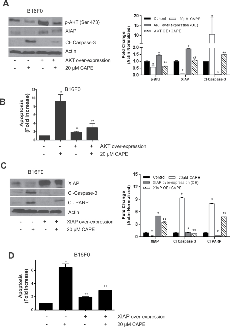
Fig. 4.
Effect of CAPE in cells overexpressing with AKT and XIAP. B16F0 melanoma cells were transiently transfected with 2 µg AKT or 2 µg of XIAP plasmid by electroporation followed by 20 µM CAPE treatment for 48 h. (A and C) Cell lysates were subjected to western blots and immunoblotted with p-AKT (ser 473), XIAP, cleaved caspase-3 and cleaved PARP. Same blots were stripped and re-probed for actin to ensure equal protein loading. (B and D) Apoptosis wasdetermined using Annexin V/FITC and propidium iodide and analyzed by flow cytometry as described in Materials and methods section. Results are expressed as mean ± SD of three independent experiments. *P < 0.05, statistically significant compared with control. **P < 0.05, statistically significant compared with CAPE treatment.

