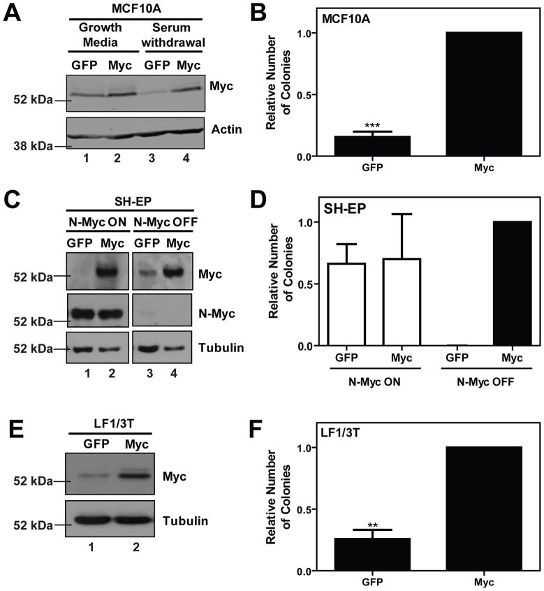
Figure 1. Human Myc promotes cellular transformation in MCF10A, SH-EP and LF1/3T human cell lines.
Control green fluorescent protein (GFP) or human Myc cDNA were introduced by infection with ecotropic, replication-incompetent retrovirus into human cell lines, MCF10A, SH-EP and LF1/3T, as described previously (Wu et al., 2004) and stable pools were isolated by fluorescence-activated cell sorting (FACS) for green fluorescent protein (GFP) expression. A) MCF10A cells (a kind gift from Senthil Muthuswamy, Ontatio Cancer Institute) were cultured as described previously (Debnath et al., 2003). Growth factor withdrawal was achieved by culturing cells in media containing 0.05% horse serum and supplemented with only 10 μL/mL insulin for one hour. Ectopic Myc expression was confirmed by immunoblotting of lysates isolated from asynchronously growing cells in full growth media and from cells subjected to 1 hour of serum and growth factor withdrawal. B) Transformation was evaluated by anchorage-independent colony growth in soft agar. Soft agar experiments were completed as previously described with the following modifications, 5 000 cells were seeded per 35 mm petri dish in triplicate, and colonies (greater than 6 cells) were counted at the end of a 2–3 week period (Oster et al., 2003). Transformation data is presented as a relative number of colonies compared to cells expressing wild-type Myc. Data represents the mean ± standard deviation from three independent experiments, **p<0.01, ***p<0.001, paired t-test. C) SH-EP Tet21/N-Myc cells (a kind gift from Manfred Schwab, German Cancer Research Center) were cultured in RPMI 1640 with 10% FBS (Breit and Schwab, 1989).
Ectopic Myc expression was confirmed by immunoblotting. This experiment was conducted both in the presence and absence of N-Myc expression. 1 μg/mL tetracycline (Sigma, St. Louis, MO) was added to growth media 48 hours prior to experiments to inactivate N-Myc expression. D) Soft agar transformation experiments were conducted as above, both in the absence (N-Myc ON) and presence (N-Myc OFF) of tetracycline. Data represents the mean ± standard deviation from two independent experiments. E) LF1/TERT/LT/ST cells (a kind gift from John Sedivy, Brown University) were grown in HAM F10 media and supplemented with 15% fetal bovine serum (FBS) (Wei et al., 2003). Ectopic Myc expression was confirmed by immunoblotting. F) Soft agar transformation experiments were conducted as above and data represents the mean ± standard deviation from three independent experiments, **p<0.01, paired t-test.

