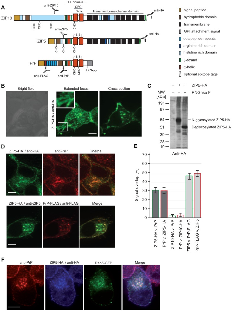
Figure 1. Co-localization of PrPC and ZIP5 at the plasma membrane and in Rab5 immunoreactive endosomes.
(A) Schematic comparing molecular features of ZIPs 5 and 10 with PrPC. (B) N2a cells were transfected with ZIP5-HA or ZIP10-HA (not shown) and the distribution of these heterologously expressed proteins analyzed by immunofluorescence. A comparison of bright field, confocal and extended focus views revealed predominant localization of ZIP5 at the plasma membrane and on vesicular structures with a negligible background staining in non-transfected cells. Both proteins can also be found in punctate structures within neuritic membrane protrusions (see insets). (C) ZIP5 is N-glycosylated within its ectodomain at multiple sites. (D) Representative co-immunofluorescence data generated with antibodies directed against endogenous PrPC and the C-terminal HA-tag present on heterologously expressed ZIP5-HA. Of note is the prominent overlap of PrPC and ZIP5-derived immunofluorescence signals; importantly, the spatial overlap of ZIP5- and PrP-dependent signals is preserved in cells which express both ZIP5-HA and PrP-FLAG when the detection is based on a polyclonal anti-ZIP5 antibody and a monoclonal anti-FLAG antibody. (E) Quantification of spatial overlap of fluorescence signals derived from PrP or PrP-FLAG with ZIP5-HA or ZIP10-HA. Split Mander's co-localization coefficients for comparing spatial signal overlaps of ZIPs and PrP were calculated in Volocity from at least 20 flattened z-stacks of individual cells. Data document mean percentage overlaps with standard error bars. (F) Co-immunofluorescence analysis of ZIP5 and PrPC with the endosomal reporter protein Rab5. This analysis was based on a previously validated Rab5-GFP expression methodology. Please note the robust co-localization of both PrPC and ZIP5 with early-endosome vesicles marked by the presence of Rab5. Scale bars are 25 µm.

