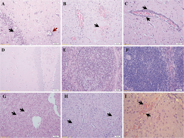
Figure 1.
Histological changes in mice infected with 7.5 × 10³ PFU of duck TMUV via the i.c. route. (A-D) Brain sections from infected mice showing (A) focal activation of glial cells (black arrow) and lymphoid perivascular cuffing (red arrow), (B) mild hemorrhage (arrow), (C) perivascular cuffing formation (arrow) and (D) non-infected controls (HE-stained). (E) Spleen sections from infected mice showing moderate lymphocyte depletion in the germinal center with the reticular structure preserved of infected mouse. (F) Spleen sections in control group. (G and H) Sections of liver and kidney showing extensive steatosis (arrow). (I) Viral antigen was detected in the cytoplasm of neural cells in the brain (arrow). (Scale bar = 20 μm).

