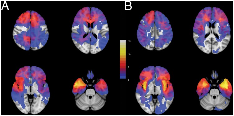
Figure 2. Glioma locations within the brain are dissimilar between cohorts.
Four transversal sections from (A) the junior team's cohort, n = 52, and (B) the senior team's cohort, n = 56, are shown superimposed on standard brain space (MNI152). More gliomas are located in the left insula and left temporal lobe in the senior team's cohort. More gliomas are located in the left supplementary motor cortex and right temporal lobe in the junior team's cohort. The legend refers to the number of patients with glioma tissue at a voxel. See Movie S1 for all transversal sections.

