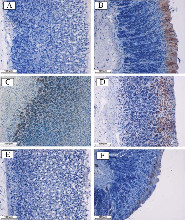
Figure 6.
Immunohistochemical analysis of Bax protein. Bax expression in the gastric tissue of rats submitted to ethanol-induced gastric mucosal lesions at different groups where (A) normal control group, (B) ulcer control group, (C) omeprazole group, (D, E, F) pre-treated group with DES at doses 5, 10 and 20 mg/kg, respectively. The antigen site appears as a brown color (IHC: 20×).

