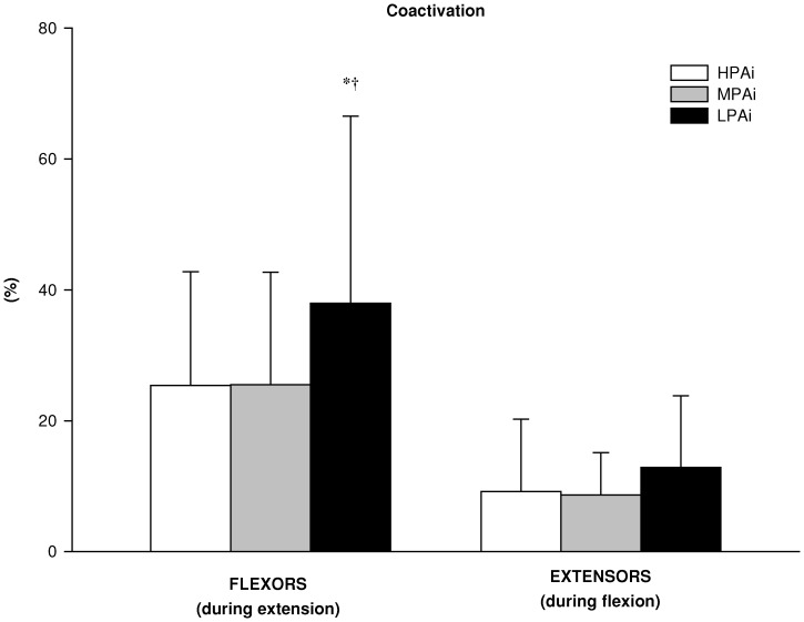
Figure 1. Neural activation in the 3 PA intensity clusters.
Percentage coactivation of the biceps femoris muscle during the knee extension maximal voluntary contraction (MVC) and of the vastus lateralis muscle during knee flexion MVC in the high (HPAi; n: 19), medium (MPAi; n: 32) and low (LPAi; n: 21) PA intensity clusters. Data are expressed as means ± SD. *significantly different from MPAi; †significantly different from HPAi.

