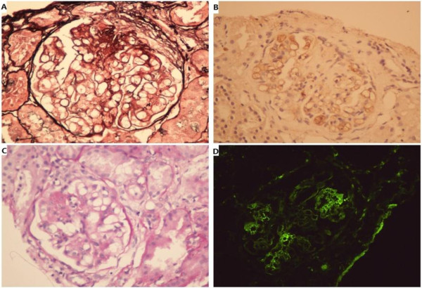
Figure 1.
Image of renal biospy. A: Focal segmental glomerulosclerosis with glomerular capillary wall collapse, balloon adhesion and fibrous small new moon body form. Surrounding the open capillary lumen, the mesangial area has no obvious proliferation. (silver staining, 400x). B: Segmental glomerular sclerosis, capillary bundle segmental collapse, balloon adhesion and cell sex small new moon body form. (PAS staining, 400x). C: HBcAg immunohistochemistry staining: HBcAg along the glomerular capillary wall and mesangial area; stage positive. (400x). D: Immunofluorescence: The glomerulus and mesangial area along the grain; sample fluorescence distribution: LgG-, LgA-, LgM++, C3-, F-, C1q-.

