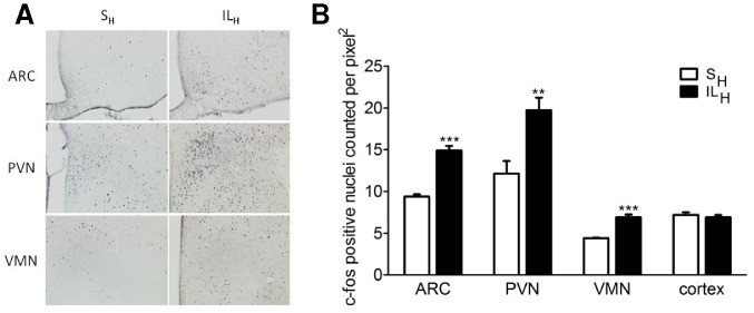
Figure 3. Immunostaining of c-fos positive cells in hypothalamic nuclei (A, B) and neuropeptides expression (C) in hypothalamus after 10 min infusion toward brain of SH or ILH.
A: photomicrographs showing c-fos positive cells localization in arcuate nucleus (ARC), paraventricular nucleus (PVN) and ventromedian nucleus (VMN) in controls (left) and ILH rats (right). B: number of c-fos-positive nuclei counted per pixel2 in controls (open bars) and ILH rats (solid bars). Values are means ±SEM; n≥6 rats/group. **p<0.01, ***p<0.001 significantly different from controls.

