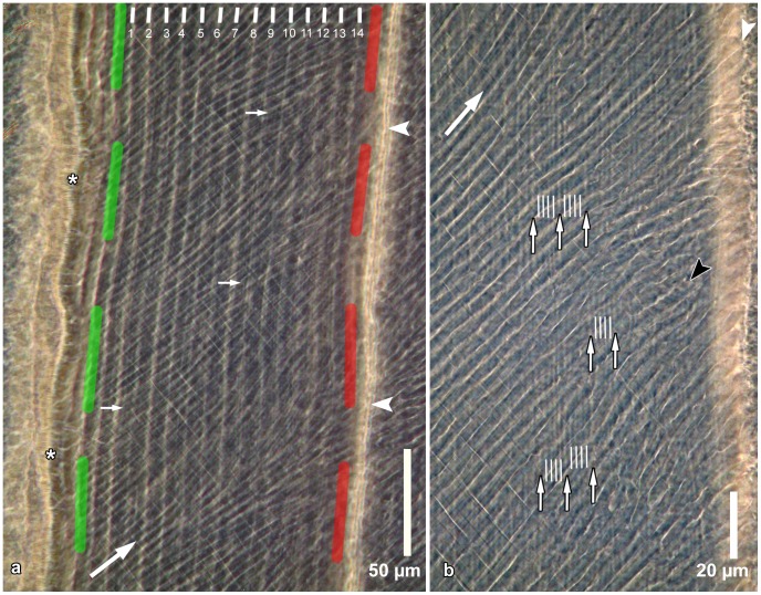
Figure 2. Incremental markings in inner enamel as seen in a ground section.
a. Enamel near the EDJ (asterisks); the depicted enamel portion is located between a calcein band (indicated by the green dashed line) and an oxytetracycline band (indicated by the red dashed line) from injections given 14 days apart. Thirteen (daily) growth increments are located between fourteen laminations (indicated by white lines). In addition, approximately half a growth increment each is located between the calcein label and lamination number 1 and between lamination number 14 and the oxytetracycline label. Arrowheads indicate the position of a Wilson band caused by the oxytetracycline injection. Large arrow indicates overall prism direction; small arrows point to finer incremental markings between successive laminations. Cuspal to top. Section viewed in transmitted light with phase contrast. b. Detail of enamel shown in Figure 2a. Five sub-daily growth increments separated by four sub-daily markings (indicated by white lines) are present between successive laminations (small arrows). Large arrow indicates overall prism direction. White arrowhead indicates Wilson band; black arrowhead indicates a prism showing sub-daily markings. Cuspal to top. Section viewed in transmitted light with phase contrast.

