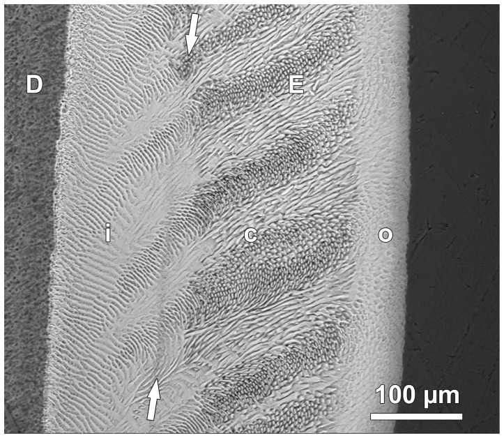
Figure 3. BSE image of etched section through lingual enamel.
The enamel (E) can be divided into three zones. The inner zone (i), adjacent to the dentin (D), is characterized by prominent interrow sheets of interprismatic enamel. The central zone (c) shows a typical Hunter-Schreger pattern with alternating bands of more or less longitudinally (parazones) and transversely cut (diazones) prisms. The outer enamel zone (o) shows a much less pronounced variation in prism orientation. Arrows mark the position of a Wilson band with a disrupted enamel structure resulting from oxytetracycline injection. Cuspal to top.

