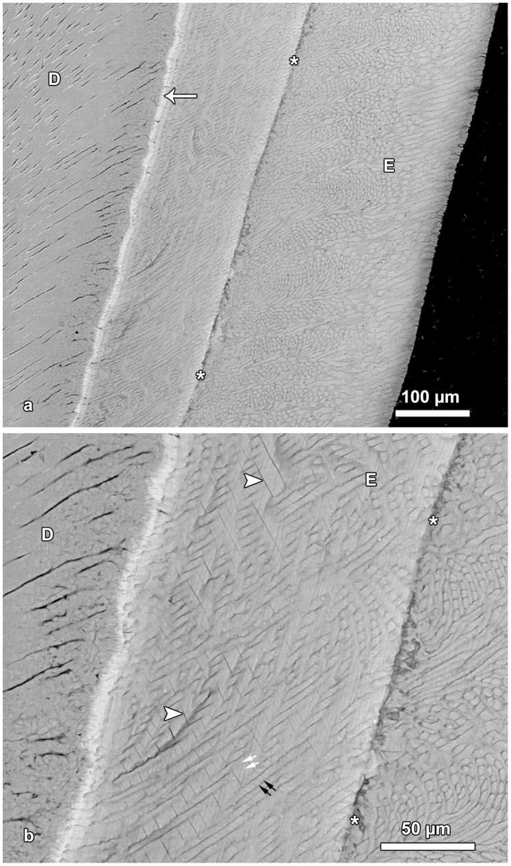
Figure 4. BSE images of hypomature lingual enamel.
a. Overview of the enamel (E) exhibiting a prominent Wilson band (asterisks) that shows a pronounced hypomineralization. Note bright line (arrow) along the EDJ, indicative of a higher mineral content. D = dentin. Cuspal to top. b. Detail of Figure 4a. A disruption of enamel microstructure is visible along the Wilson band (asterisks). Note presence of fine incremental markings in the interprismatic enamel (white arrows) and in the enamel prisms (black arrows). Arrowheads mark fine clefts in the enamel (E); D = dentin.

