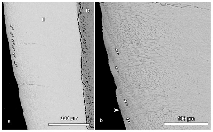
Figure 5. BSE images illustrating laminations in outer enamel.
a. Regularly spaced laminations (arrows) are visible in the outer portion of the enamel (E). In contrast, no laminations are discernible in central and inner enamel. D = dentin. Cuspal to top. b. Detail of Figure 5a. Laminations (arrows) in outer enamel. Note that the outcrop of a lamination on the OES is mostly associated with the presence of a small trough (furrow) on the surface. However, single laminations reach the enamel surface without producing a trough (arrowhead). Cuspal to top.

