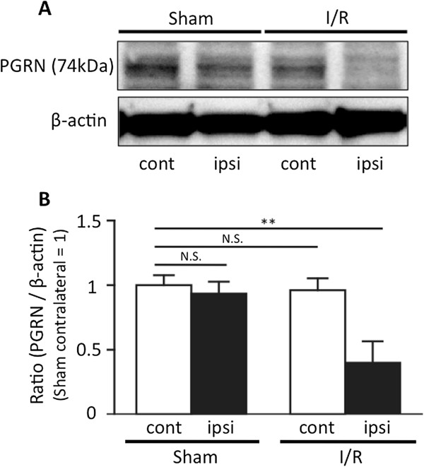
Figure 1.

Expression of progranulin in the ischemia-reperfusion injured brain. Progranulin (PGRN) expression was significantly decreased following ischemia-reperfusion (I/R) insults. (A) Representative PGRN bands from the Western blotting analysis of brain tissue taken from sham-operated and I/R animals; ipsilateral and contralateral hemispheres to the middle cerebral artery occlusion (MCAO). (B) Optical densitometry quantification of PGRN protein levels, normalized to β-actin. In the I/R brain, the expression of PGRN was significantly decreased 24 h after the induction of transient cerebral ischemia. **P <0.01 vs. sham contralateral brain; one-way ANOVA followed by Dunnett's test; n = 4 for each group.
