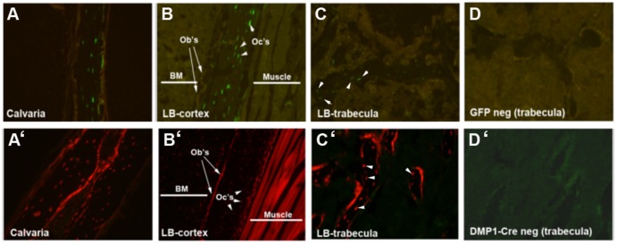
Figure 1. Histology of Dmp1-GFP and Dmp1Cre/Ai9 bones.
Detection of GFP expression in Dmp1-GFP derived histological sections (A–D). A specific GFP signal was localized in osteocytes (arrowheads) of cortical bone in calvaria (A) and femur (B) and in trabeculae (C). Detection of tdTomato expression in histological sections derived from Dmp1Cre/Ai9 transgenic mice (A’–D’). Cre-directed recombination was detected in osteocytes (arrowheads) and in the osteoblast layer (arrows) of cortical bone from calvaria (A’) and femur (B’) and trabecular bone (C’). tdTomato expression was also present in skeletal muscle (B’). Sections derived from GFP negative (D) and Dmp1Cre negative (D’) mice served as controls. Images were taken at 10X magnification and are representative of histology performed on 6 mice of each genotype. BM, bone marrow; LB, long bone; Ob’s, osteoblasts; Oc’s, osteocytes.

