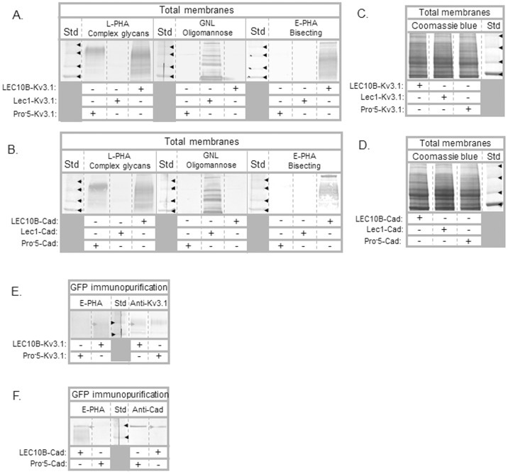
Figure 2. Lectin blots of total membranes and immunopurified Kv3.1 and E-cadherin proteins from transfected CHO cell lines.
Total membranes (∼25 µg) from Pro-5, Lec1, and LEC10B cells transfected with wild type Kv3.1 (A) and E-cadherin (B) were probed with L-PHA (∼5 µg/mL), E-PHA (5–10 µg/mL), and GNL (∼10 µg/mL). Similar amounts of electrophoresed proteins from total membranes were also stained with Coomassie blue (C,D). Black arrowheads denote the 75, 100, 150 and 250 kDa markers. Lectin blots of immunopurified GFP tagged Kv3.1 and E-cadherin from transfected Pro-5 and LEC10B cells (E,F). Glycoproteins were probed with E-PHA (5–20 µg/mL). Western blots were run in parallel to denote position and relative amount of GFP-Kv3.1 and E-cadherin protein. Grey arrowheads point to GFP tagged Kv3.1 (E) and E-cadherin (F) proteins expressed in LEC10B cells while black arrowheads represent the 100 and 150 kDa markers.

