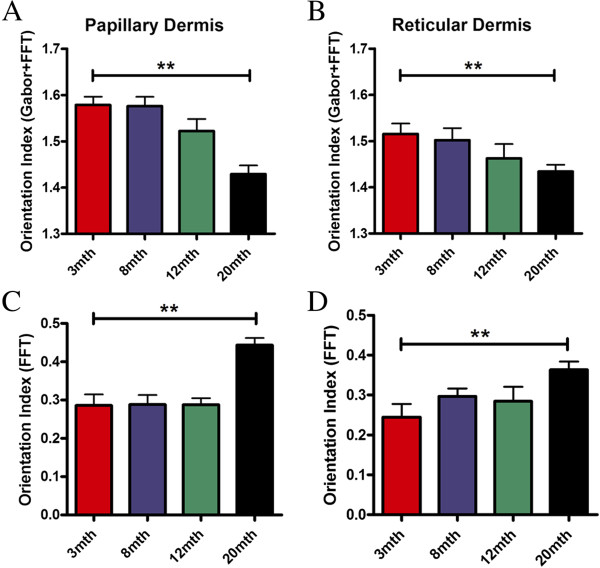
Figure 4.
Increasing age corresponds with differential patterns of decline in collagen organisation in the different layers of the dermis. A progressive decline in integrity was seen in both papillary and reticular compartments by our method (A and B). While FFT alone was able to detect changes by 20mth, there was no evidence of progressive decline in either compartment (C and D). The bar equates to the mean and hair lines are standard error of the mean (S.E.M.). One way ANOVA with post-hoc Dunnett’s test compared to 3mth group was performed, for all tests: ** p < 0.01 (n > 3 animals per group).

